Histology(Ch1-5)
Where did Hematoxylin come from
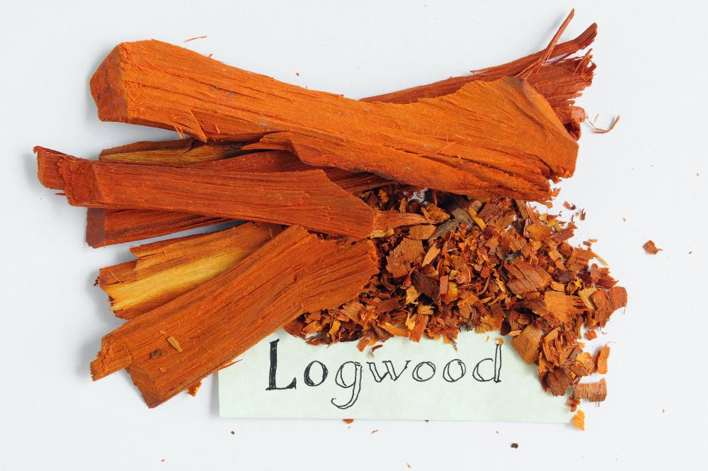
From Logwood
Is hematoxylin an acid or a base
Base
What does Hematoxylin stain
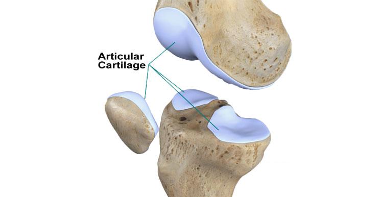
RNA, DNA, Carbohydrates and cartilage, because they're acidic
What does eosin stain
Most proteins and some extracellular fibers
Is eosin an acid or a base
Negative Acid
What color does Hematoxylin stain the Nucleus
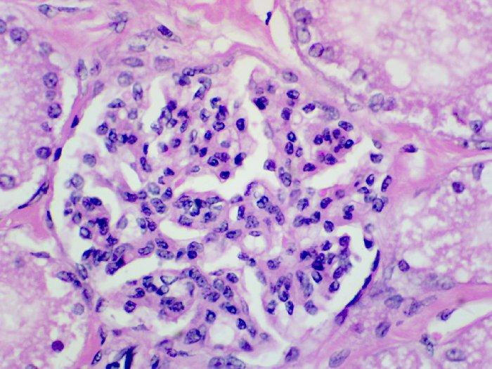
Purplish blue
What color does eosin stain
Red or pink
What are the compounds in Masson's trichrome

Hematoxylin, acid fuchsin, methyl blue
What color does Masson's trichrome stain Nuclei
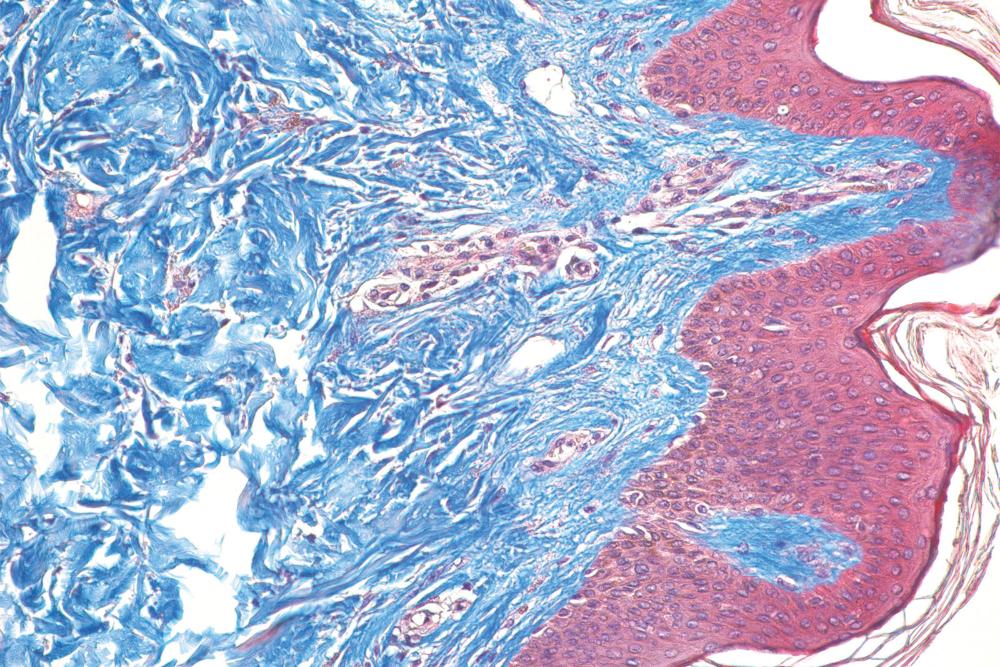
Blue
What color does Masson's trichrome stain connective tissue and basement layers
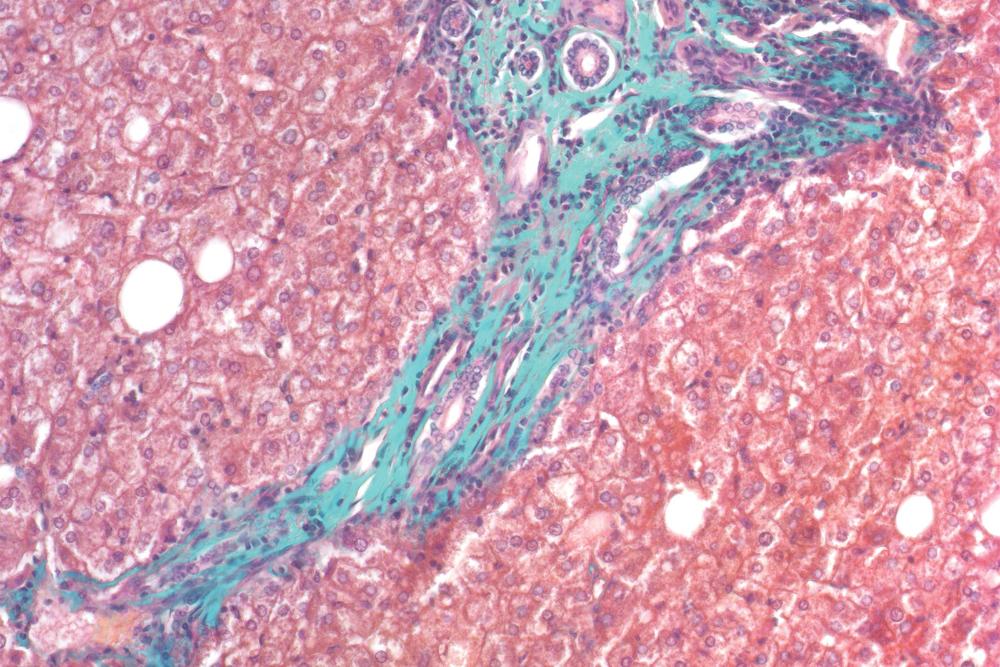
Green
Whats Van Gieson stain used for?
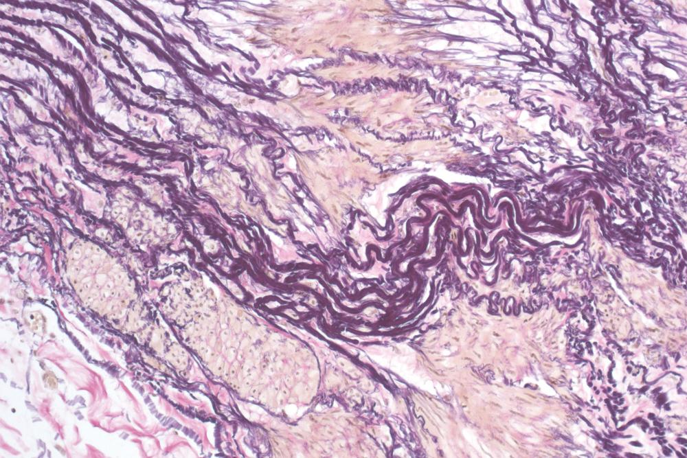
For connective tissue detection
Whats Geimsa stains used for
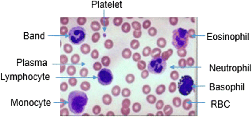
Detecting Erythrocytes in the bone marrow to see if they're sick, and also for blood smears (is a test used to see the shape, size and appearance of red blood cells)
What color does giemsa stains stain red blood cells
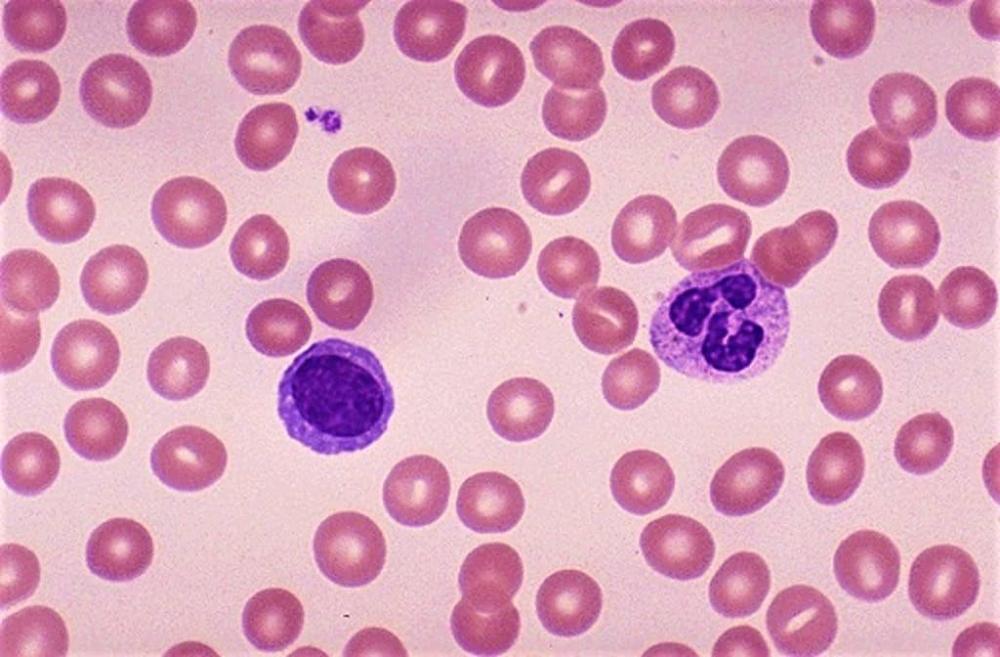
Pink
What color does giemsa stains stain white blood cells

Dark purple/ blue
What does silver stain used for
To stain nerves
Whats Golgi-Cox used for
To stain nerves
What are the compounda in Golgi-Cox

Mercuric chloride, potassium chromate K2CrO4, potassium dichromate K2Cr2O7
Whats Cresyl violet used for
Detecting the endoplasmatic reticulum of the body of nerve cells(perikarya)
What color does Cresyl Violet stain body of neurons?
Brown
Name lipid stains

Nile Blue, Oil Red O and Sudan black
How thick is sectioning if done with microtome
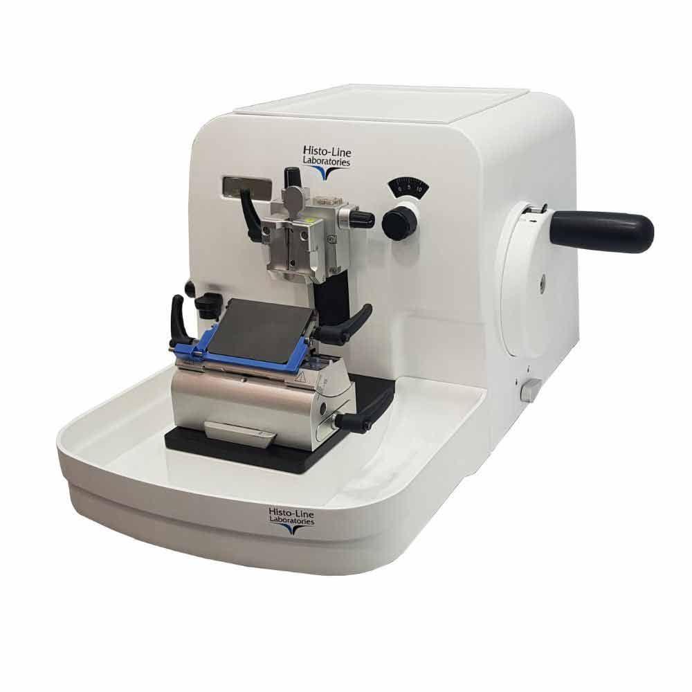
10-20 Microns
What are low power Lenses

*1.6, *5 and *10
What are Hight low power Lenses

*20, *40, *63 and *100
What is the wave length range for halogen lamp(tungsten lamp)
starts at 300-400nm to 700-800nm
What does the magnification of Light microscope depend on?

the Magnification of the eye piece and Objective lens, for example if you had a eyepiece in a light microscope that can magnify by x10 and then objective lens that can magnify by x10 then in total your light microscope can magnify by x100
Whats the best resolution obtainable from a light microscope
0.2 Microns or 200nm
How big are cells in diameter
20-40 microns
Whats the Wave length in a Electron microscope
0.004nm
What does electro microscope use instead of lenses to focus the light
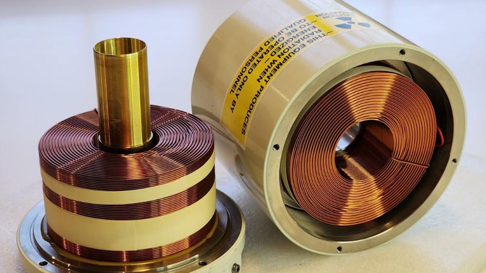
electromagnetic coils
What is the Numerical aperture for electron microscope
0.012
What is the greatest magnification for electron microscope
x50,000
How thick are the prepared sections for Electron microscope
60-100nm
How thick is the plasmatic membrane
8-10nm
How much is the diameter of the Nucleus
10 Microns
What is the function of the Nucleolus
Making Ribosomes
How much of cell volume do endoplasmic reticulum constitute
10%
What is the function of the endoplasmic reticulum
- Store Calcium
- make membranes for the other organelles(rough: makes the necessary protein, Smooth: makes the necessary lipids)
How many membranes does the Smooth ER have
it has the same structure as the plasmatic membrane, a single lipid bilayer membrane
How wide are the lumens of Rough ER
20-30nm
How wide are the lumens of Smooth ER
30-60nm
What is Cisternae
its the first 3-7 discs of the Golgi apparatus, the discs that receive, modify, transform and pack proteins
What is the trans face of Golgi apparatus
its the face that transports the proteins to the plasmatic membrane
What is the function of secreting vesicles
to get molecules to the plasmatic membrane
What is the function of Endosomes
they bring molecules from outside the cell inward, then to the lysosomes so that it can break it down
What is the function of Macropinosomes
they bring large amounts of fluid into the cell
What is the function of endosomes that do Phagocytosis
they kill bacteria
What is the function of Peroxisomes
they degrade fatty acids by adding an oxygen, then convert them to cholesterol
Where are Peroxisomes common/abundant

liver and kidney
How big are vesicles
50-200nm
What is Christae
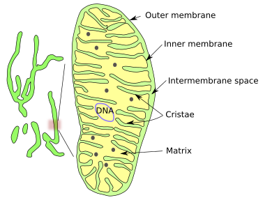
they're the folds of the inner membrane of mitochondria that folds inward, the folds are empty intermediate space
What is the function of Actin filaments
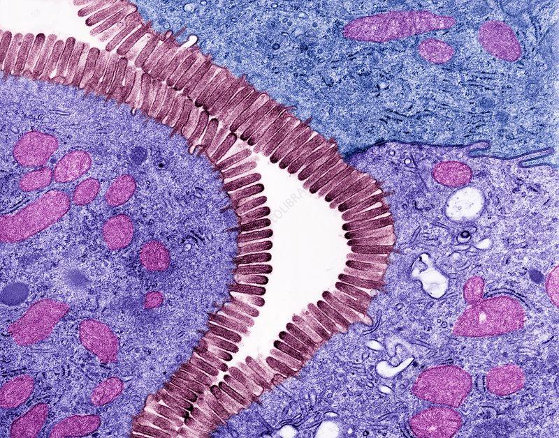
- Act as a track for Myosin
- they link cells to each other by tight or adhesion junctions
- they're an important component in the creation of cell protuberances(elevations on the surface of the cell membrane) like Microvilli in the intestine
How thick are actin filaments
7nm
What is the function of Intermediate filaments
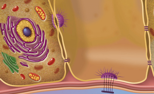
they make it easier for cells to stick to each other by desmosomes and hemidesmosomes
What is the function of Microtubules
- act as tracks for motors (kinesins, dynein) that move vesicles around the cell
- they make the flagella and cilia
- they make the mitotic and meiotic spindles
What makes Microtubules
Centrosomes
How big are Intermediate filaments
10nm
How big are Microtubules
25nm
What are the things you do in tissue processing for electron microscope that you don't do in light Microscopy
1)The fixative isn't formality It's rather glutaraldehyde
2)Post fixation(another fixation after glutaraldehyde) by osmic acid
3)We put the tissue in propylene oxide that allows us to impregnate it later with resin instead of wax
4) Machine For sectioning is Ultramicrotome That has a glass Or diamond knife
5)Stains are heavy salt Metals like Osmium, uranyl acetate And lead To increase. The contrast Because they dispersed
Why do we post-fixate with osmic acid in tissue processing for electron microscopy
glutaraldehyde results in cross-linking proteins that reserves protein structure while osmic acid preserves lipids