Instructions for Side by Side Printing
- Print the notecards
- Fold each page in half along the solid vertical line
- Cut out the notecards by cutting along each horizontal dotted line
- Optional: Glue, tape or staple the ends of each notecard together
Anatomy & Physiology: The Integumentary System
front 1 Integument | back 1 -makes up 16% of your body weight
|
front 2 What are the 2 components of the cutaneous membrane? | back 2  1. Epidermis
|
front 3 Epidermis | back 3  -superficial epithelium
|
front 4 Dermis | back 4 -underlying area of connective tissue |
front 5 Accessory Structures | back 5  -hair, exocrine glands, and nails
|
front 6 Hypodermis | back 6  -aka superficial fascia or subcutaneous layer
|
front 7 Functions of the Integumentary System | back 7  -protection of underlying tissues & organs
|
front 8 4 layers of thin skin | back 8 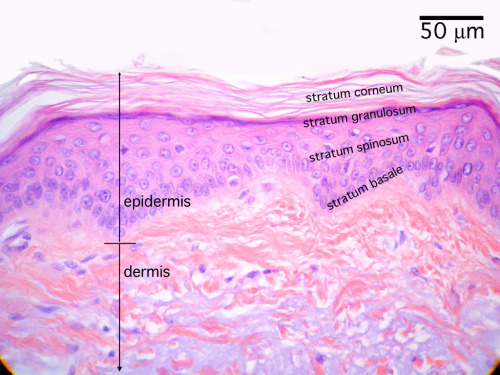 1.Stratum Corneum
|
front 9 5 Layers of thick skin | back 9  1.Stratum Corneum
|
front 10 Stratum Basale | back 10  -aka stratum germinativum
|
front 11 Basal Cells | back 11 -cells that divide to replace the more superficial keratinocytes that are shed at the epithelial surface |
front 12 Stratum Spinosum "Spiny Layer" | back 12  -consists of 8-10 layers of keratinocytes bound together by desmosomes
|
front 13 Dendritic Cells | back 13  -aka Langerhans cells
|
front 14 Stratum Granulosum "Grainy Layer" | back 14 %20stratification.jpg) -consists of 3-5 layers of keratinocytes
|
front 15 Keratin | back 15 -A tough, fibrous protein
|
front 16 Stratum Lucidum "Clear Layer" | back 16  -only in thick skin
|
front 17 Stratum Corneum | back 17 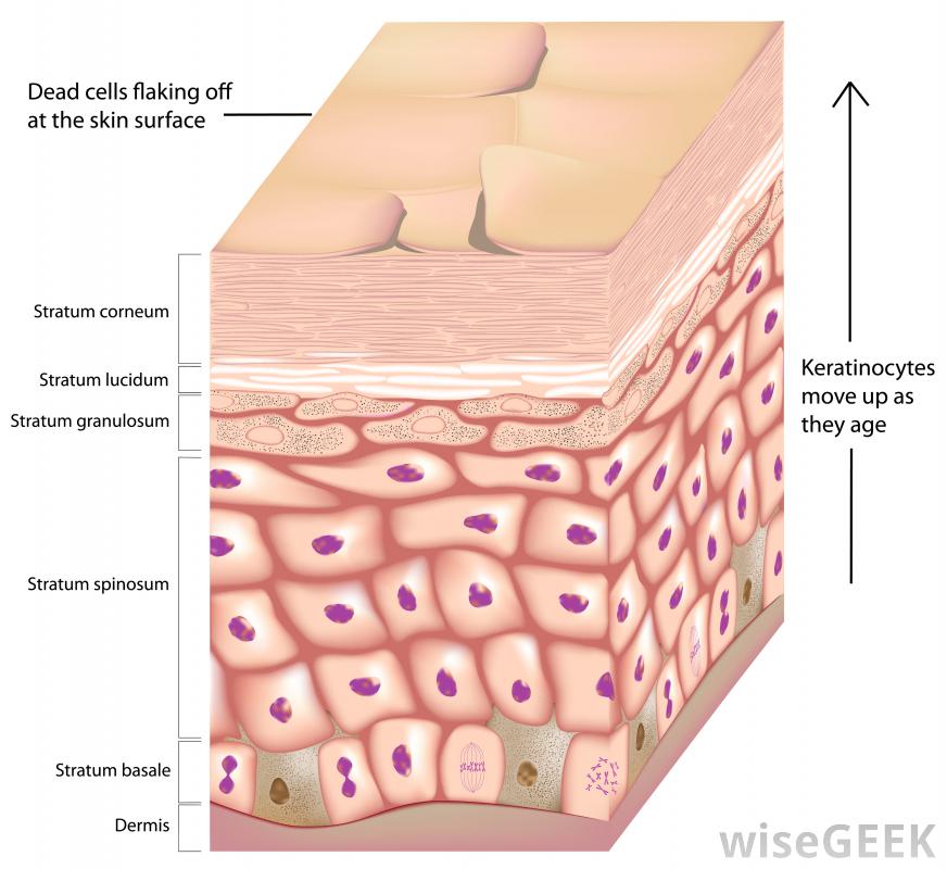 -exposed surface
|
front 18 Keratinization | back 18 -the formation of protective, superficial layers of cells filled with keratin
|
front 19 How long does it take for a cell to move from the Stratum Basale to the Stratum Corneum? | back 19  Around 7-10 days |
front 20 Insensible Perspiration | back 20 -water from interstitial fluids slowly penetrates to the surface and evaporates into the air
|
front 21 Sensible Perspiration | back 21 -produced by active sweat glands
|
front 22 What are the two pigments found in the epidermis? | back 22 Carotene and Melanin |
front 23 Carotene | back 23 An orange-yellow pigment that normally accumulates in epidermal cells
|
front 24 Melanin | back 24  pigment produced by the melanocytes
|
front 25 Melanocytes | back 25 -manufacture both types of melanin from the amino acid tyrosine and packages it into intracellular vesicles called melanosomes |
front 26 The __________ in keratinocytes protects your epidermis and dermis from the harmful effects of sunlight, which contains significant amounts of ultraviolet radiation (UV). | back 26 melanin |
front 27 Blood contains red blood cells filled with the pigment ___________, which binds and transports oxygen in the bloodstream. | back 27 hemoglobin |
front 28 Cyanosis | back 28 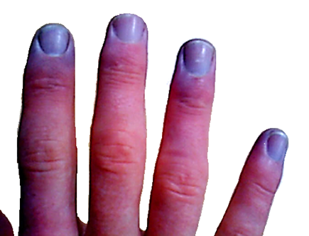 when the skins turns blue
|