Instructions for Side by Side Printing
- Print the notecards
- Fold each page in half along the solid vertical line
- Cut out the notecards by cutting along each horizontal dotted line
- Optional: Glue, tape or staple the ends of each notecard together
muscles of the lower leg
front 1 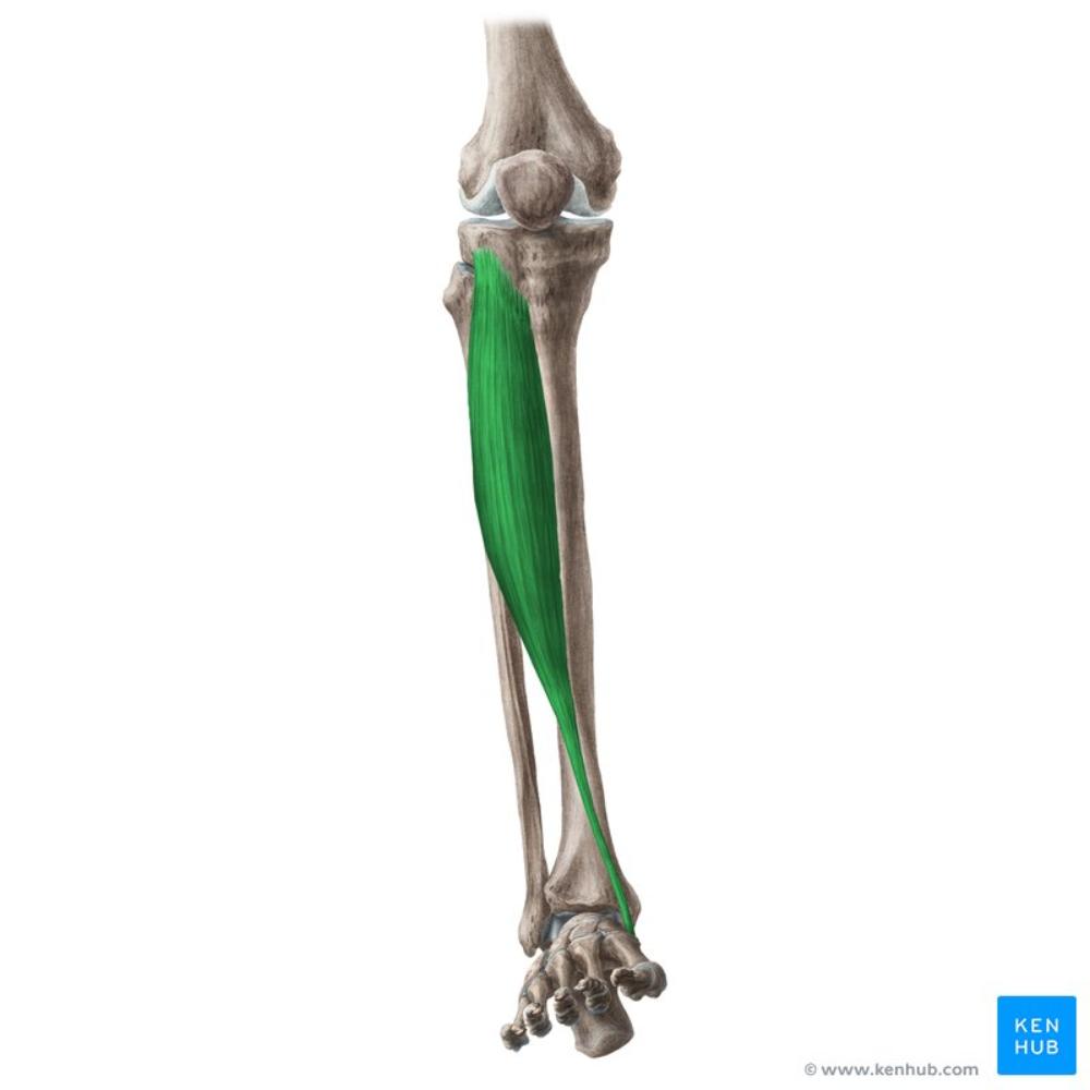 tibialis anterior | back 1 O: Lateral condyle of tibia, upper ½ of lateral surface of tibia, interosseous membrane I: medial and plantar surface of middle cuneiform, base of 1st metatarsal A: dorsiflexion, inversion (supination) of the foot N: deep peroneal |
front 2 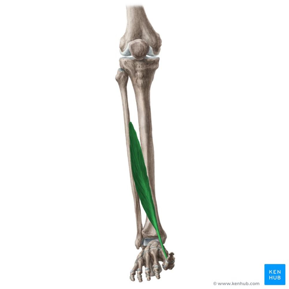 Extensor hallicus longus | back 2 O: middle half of anterior fibula, interosseous membrane I: base of distal phalanx of great toe A: extends & hyperextends great toe, dorsiflexion & inverts foot N: deep peroneal |
front 3 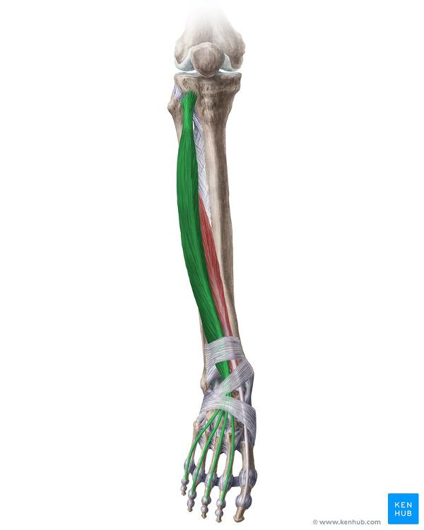 Extensor digitorum longus | back 3 O: upper 2/3 of anterior fibula, interosseous membrane, lateral condyle of fibula I: dorsal surface of lateral 4 toes, base of middle and distal phalanges A: extends toe, dorsiflexion and eversion of foot N: deep peroneal |
front 4 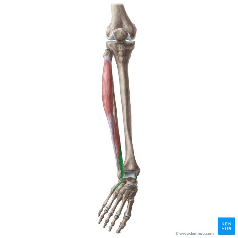 peroneus (fibularis) tertius | back 4 O: lower 2/3 of the fibula; interosseous membrane I: dorsal surface of the base of 5th metatarsal A: dorsiflexion and eversion of foot N: deep peroneal |
front 5 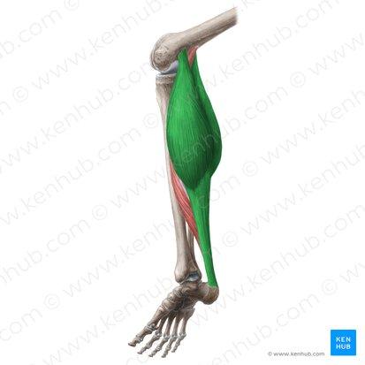 gastrocnemius | back 5 O: lateral head: lateral condyle and posterior surface of femur medial head: popliteal surface of femur above medial condyle I: posterior surface of calcaneus through achillies tendon A: plantarflexion of foot, flexes leg at knee N: tibial nerve |
front 6 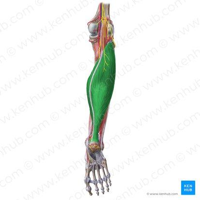 soleus | back 6 O: Posterior surface of the tibia, upper 1/3 of I: posterior surface of calcaneus via the achillies tendon A: plantarflexion of the foot N: tibial nerve |
front 7 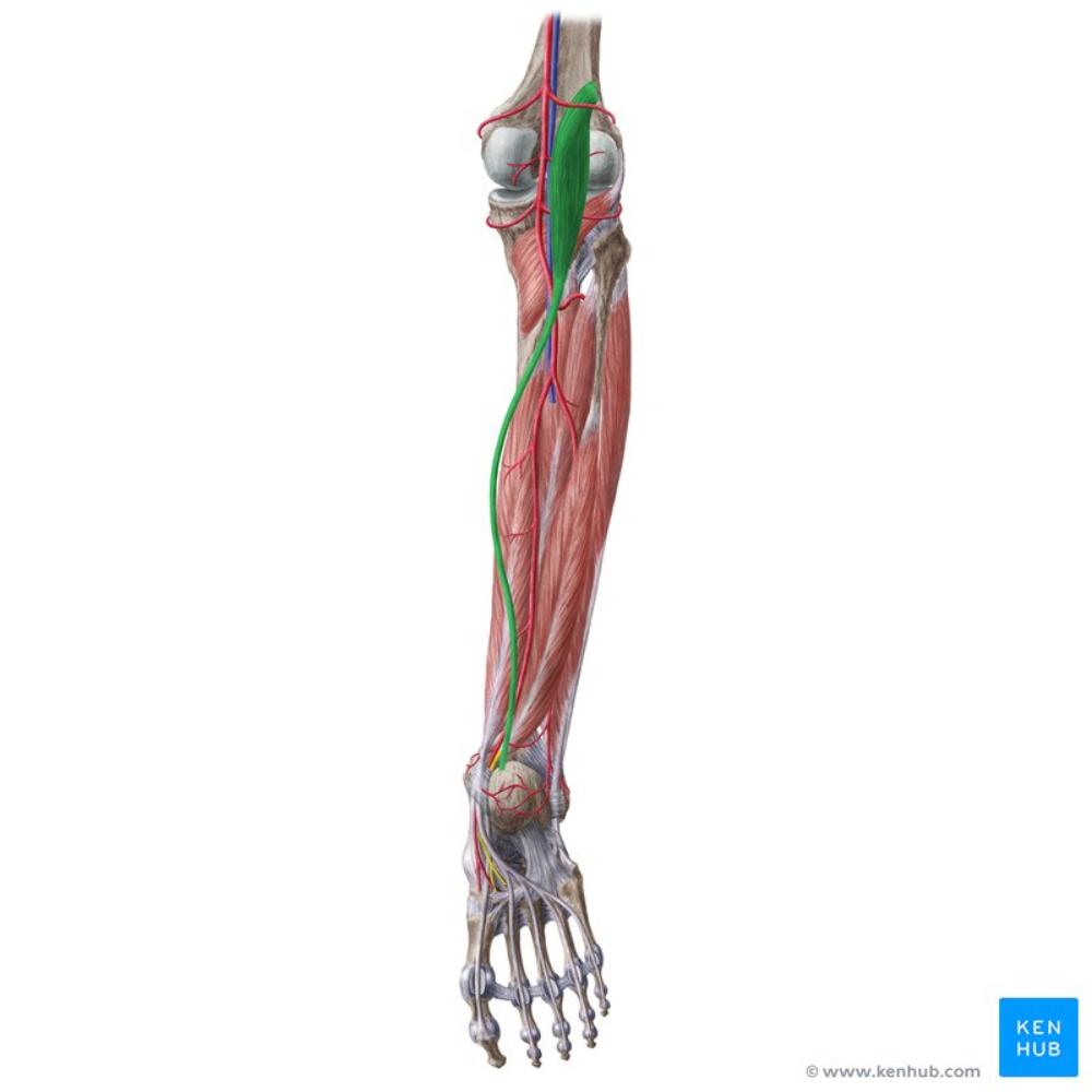 plantaris | back 7 O: lateral supracondylar ridge of femur, oblique popliteal ligament I: posterior surface of calcaneus A: plantarflexion of foot, flexes leg at knee N: tibial nerve |
front 8 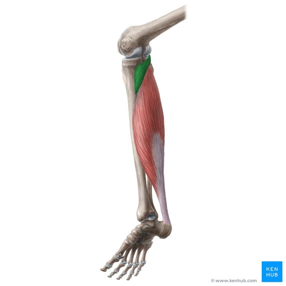 popliteus | back 8 O: lateral surface of the lateral condyle of the femur I: upper part of the posterior surface of the tibia A: medial rotation of the leg, flexion of the leg N: tibial nerve |
front 9 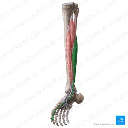 flexor hallucis longus | back 9 O: Lower 2/3 of posterior surface of the shaft of I: base of distal phalanx of the great toe A: flexion of great toe (distal phalanx), assist PF and inversion of the foot N: tibial nerve |
front 10 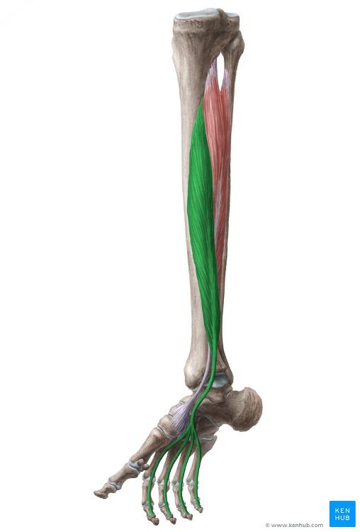 flexor digitorum longus | back 10 O: medial part of posterior surface of the tibia I: base of 2-5 distal phalanges A: flexion of 2-5 distal phalanges, assist PF and inversion of foot N: tibial nerve |
front 11 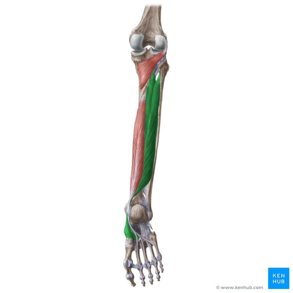 tibialis posterior | back 11 O: lateral part of the posterior surface of the tibia, proximal 1/2 of the posterior surface of the fibula I: navicular tuberosity, cuboid, cuneiforms, 2-4 metatarsals, sustentaculum tali A: flexion of 2-5 distal phalanges, assist PF and inversion of the foot N: tibial nerve |
front 12 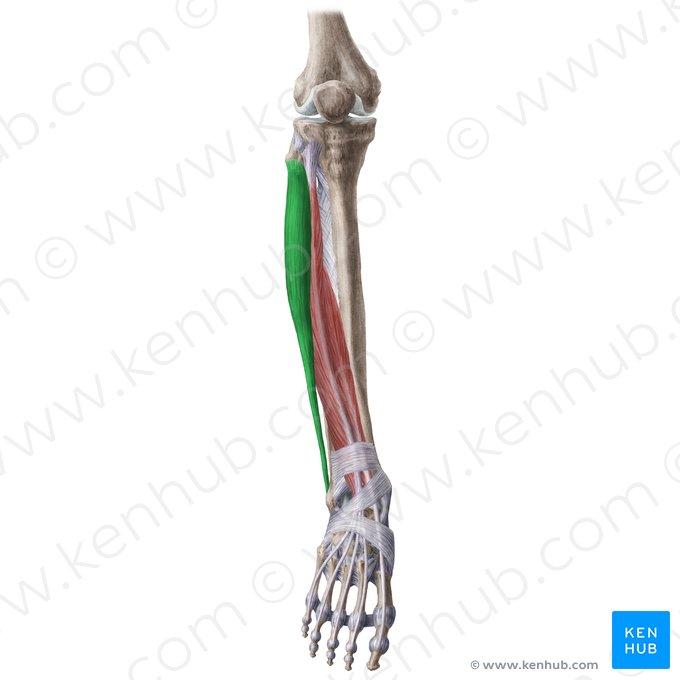 peroneus (fibularis) longus | back 12 O: upper 2/3 of the lateral surface of the fibula I: lateral surface of medial cuneiform, base of 1st metatarsal A: plantarflexion & eversion of foot N: superficial peroneal nerve |
front 13 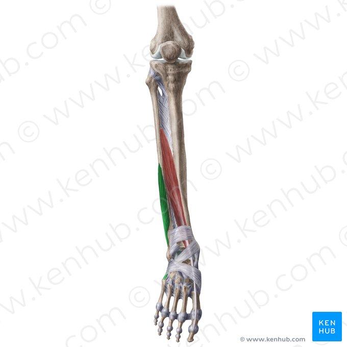 peroneus (fibularis) brevis | back 13 O: lower 2/3 surface of the fibula I: lateral side of the base of 5th metatarsal A: plantarflexion & eversion of foot I: superficial peroneal nerve |