Instructions for Side by Side Printing
- Print the notecards
- Fold each page in half along the solid vertical line
- Cut out the notecards by cutting along each horizontal dotted line
- Optional: Glue, tape or staple the ends of each notecard together
Radiographic positioning review (ALL)
front 1 Image Receptor Size: 10 x 12 | back 1  AP of the Knee |
front 2 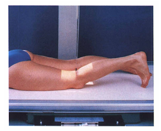 Image Receptor Size: 10 x 12 | back 2  PA of the Knee |
front 3 Image Receptor Size: 10 x 12 | back 3  Lateral of the Knee, Mediolateral |
front 4 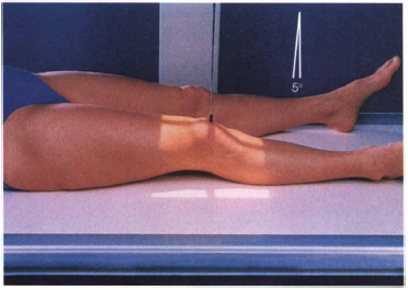 Image Receptor Size: 10 x 12 | back 4  AP Oblique of the knee, Lateral rotation |
front 5  Image Receptor Size: 10 x 12 | back 5  AP Oblique of the knee, Medial rotation |
front 6  Image Receptor Size: 10 x 12 | back 6  AP of the knee, Weightbearing |
front 7 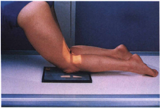 Image Receptor Size: 8 x 10 | back 7  PA axial of the intercondyloid fossa, Holmbland Method |
front 8 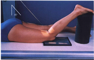 Image Receptor Size: 8 x 10 | back 8  PA Axial of the intercondyloid fossa, Camp-Coventry Method, Tunnel View |
front 9  Image Receptor Size: 8 x 10 | back 9  AP Axial of the intercondyloid fossa, Beclere Method, Tunnel view |
front 10 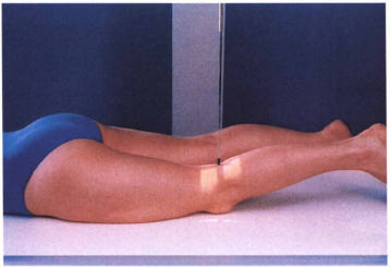 Image Receptor Size: 8 x 10 | back 10  PA patella |
front 11 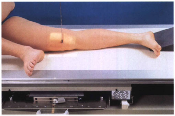 Image Receptor Size: 8 x 10 | back 11  Lateral patella |
front 12  Image Receptor Size: 8 x 10 | back 12 Tangential patella, Sattegast Method, Sunrise View |
front 13  Image Receptor Size: 8 x 10 | back 13 Tangential patella, Hughston Method |
front 14  Image receptor Size: 10 x 12 | back 14 Tangential patella, Merchant Method |
front 15 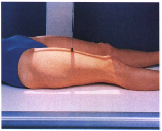 Image Receptor Size: 14 x 17 | back 15 AP of the femur (distal) |
front 16 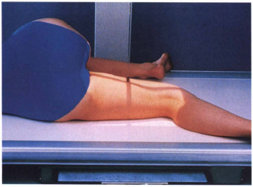 Image Receptor Size: 14 x 17 | back 16 Lateral of the femur, Mediolateral (distal) |
front 17 Image Receptor Size: 14 x 17 | back 17 AP of the Pelvis |
front 18  Image Receptor Size: 14 x 17 | back 18 AP Oblique of the Pelvis, Modified Cleaves Method, Frogleg (Bilateral) |
front 19  Image receptor Size: 10 x 12 | back 19 AP Axial of the Pelvis, Outlet, Taylor Method |
front 20  Image Receptor Size: 8 x 10 | back 20 AP Axial of the pelvis, Inlet, Bridgeman Method |
front 21  Image Receptor Size: 10 x 12 Lengthwise | back 21 AP of the Hip |
front 22  Image Receptor Size: 10 x 12 Lengthwise or crosswise | back 22 Lateral of the hip, Mediolateral, Lauenstein & Hickey Methods, Frogleg |
front 23  Image Receptor Size: 10 x 12 with grid Lengthwise | back 23 Axiolateral of the hip, Danelius-Miller, Cross-table |
front 24  Image Receptor Size: 10 x 12 (24 X 30) | back 24 Modified Axiolateral of hip, Clements-Nakayama Modification |
front 25  Image Receptor Size: 10 x 12 | back 25 AP Oblique of hip, Judet Method |
front 26  Image receptor Size: 10 x 12 Crosswise | back 26 AP of the clavicle |
front 27  Image Receptor Size: 10 x 12 Crosswise | back 27 AP axial of the clavicle |
front 28  Image Receptor Size: 10 x 12 | back 28 AP of the shoulder, External rotation |
front 29 Image Receptor Size: 10 x 12 Crosswise or Lengthwise | back 29 AP of the shoulder, Neutral rotation |
front 30  Image Receptor Size: 10 x 12 | back 30 AP of the shoulder, Internal rotation |
front 31  Image Receptor Size: 10 x 12 Lengthwise | back 31 Transthoracic Lateral of the shoulder, Lawrence Method |
front 32 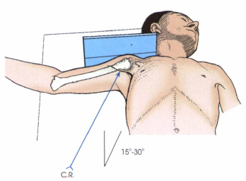 Image Receptor Size: 8 x 10 | back 32 Inferosuperior Axial of the shoulder, Lawrence Method, Axillary |
front 33  Image Receptor Size: 10 x 12 | back 33 AP Oblique of the shoulder, Grashey Method |
front 34 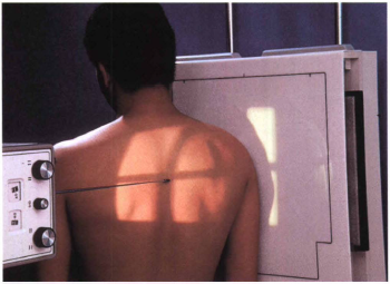 Image Receptor Size: 10 x 12 | back 34 PA Oblique of the shoulder, Scapular Y |
front 35  Image Receptor Size: 14 x 17 Crosswise | back 35 PA of the ribs, Upper Anterior Ribs |
front 36 Image Receptor Size: 14 x 17 | back 36 PA of the Ribs, Lower Anterior Ribs |
front 37  Image Receptor Size: 14 x 17 | back 37 AP of the ribs, Upper Posterior Ribs |
front 38  Image Receptor Size: 14 x 17 Crosswise | back 38 AP of the Ribs, Lower Posterior Ribs |
front 39  Image Receptor Size: 14 x 17 Lengthwise | back 39 AP Oblique of the Ribs, RPO |
front 40  Image Receptor Size: 14 x 17 Lengthwise | back 40 AP Oblique of the Ribs, LPO |
front 41  Image Receptor Size: 8 x 10 Lengthwise | back 41 AP Axial of the Cervical spine |
front 42  Image Receptor Size: 8 x 10 Lengthwise | back 42 AP of the Cervical Vertebrae, Open Mouth, For Atlas & Axis |
front 43  Image Receptor Size: 8 x 10 Lengthwise | back 43 Lateral R or L, Grandy Method |
front 44  Image Receptor Size: 8 x 10 Lengthwise | back 44 Lateral R or L, Hyperflexion/Hyperextension |
front 45  Image Receptor Size: 8 x 10 | back 45 AP Axial Oblique of Cervical vertebrae, RPO |
front 46  Image Receptor Size: 8 x 10 | back 46 AP Axial Oblique of Cervical vertebrae, LPO |
front 47 Always perform a cross-table lateral cervical spine for severe injury
FIRST. Do not move the patient or remove cervical
collars. | back 47 Trauma (of the cervical vertebrae) |
front 48  Image Receptor Size: 8 x 10 | back 48 AP of the cervical vertebrae, Fuchs Method |
front 49  Image Receptor Size: 10 x 12 | back 49 Lateral of the cervicothoracic region (R or L), Swimmers |
front 50  Image Receptor Size: 8 x 10 or 10 x 12 Lengthwise | back 50 AP Axial of the cervicothoracic region, Vertebral Arch, Pillars |
front 51  Image Receptor Size: 14 x 17 or 7 x 17 Lengthwise | back 51 AP of thoracic spine |
front 52  Image Receptor Size: 14 x 17 or 7 x 17 | back 52 Lateral of thoracic vertebrae (R or L) |
front 53  Image Receptor Size: 14 x 17 or 11 x 14 Lengthwise | back 53 AP of the lumbar spine |
front 54  Image Receptor Size: 14 x 17 or 11 x 14 | back 54 Lateral of the lumbar spine (R or L) |
front 55  Image Receptor Size: 8 x 10 Lengthwise | back 55  (L5 - S1) Lumbosacral Junction, Lateral of the lumbar spine (R or L) |
front 56  Image Receptor Size: 8 x 10 or 10 x 12 | back 56 AP Oblique of the lumbar spine, (RPO/LPO) |
front 57  Image Receptor Size: 14 x 17 Lengthwise | back 57 Lateral of the lumbar vertebrae, Weightbearing Method, Flexion |
front 58  Image Receptor Size: 14 x 17 Lengthwise | back 58 Lateral of the lumbar vertebrae, Weightbearing Method, Extension |
front 59  Image Receptor Size: 14 x 36 | back 59 Scoliosis series, PA of the lumbar spine, Ferguson Method |
front 60  Image Receptor Size: 8 x 10 or 10 x 12 | back 60 AP Axial of the sacroiliac joints, Ferguson Method |
front 61  Image Receptor Size: 8 x 10 or 10 x 12 | back 61 AP Oblique of sacroiliac joints(RPO/LPO) |
front 62  Image Receptor Size: 10 x 12 (Sacrum) | back 62 AP Axial of the Sacrum |
front 63  Image Receptor Size: 10 x 12 (Sacrum) 8x 10 (Coccyx) | back 63 AP Axial of the Coccyx |
front 64  Image Receptor Size: 10 x 12 (Sacrum) 8x 10 (Coccyx)
Lengthwise | back 64 Lateral of Sacrum and Coccyx (R or L) |