What are the main objectives of root canal procedures are:
- ___
- ___
- adequate enlargement, shaping, cleaning, and disinfection of all pulpal spaces
- obturation of these spaces with an acceptable filling material
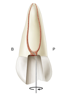
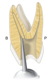
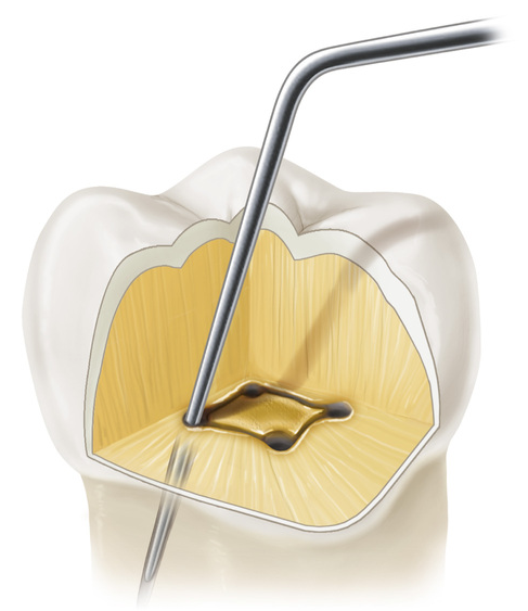
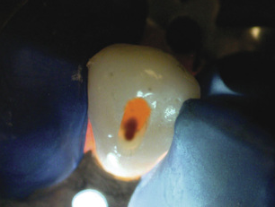
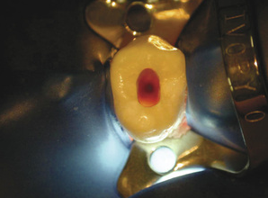
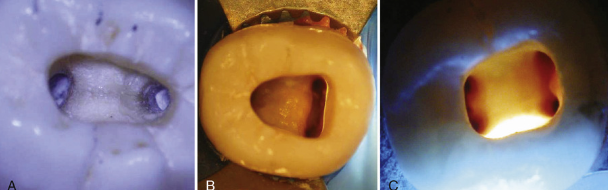
Allowing ___ to remain in the pulp chamber may help locate a calcified root canal orifice. Tiny bubbles may appear in the solution, indicating the position of the orifice.
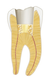
sodium hypochlorite (NaOCl)
Specifically, the use of the ___, which is intended to provide superior magnification, increased lighting, and enhanced visibility, is recommended to determine the location of root canal orifices in the properly prepared coronal access.
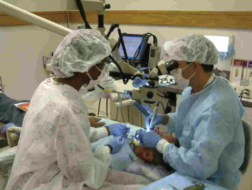
dental operating microscope (DOM)
Additional studies have noted that use of the DOM improves the detection of mesiopalatal canals to more than ___% in maxillary first molars and ___% in maxillary second molars.
90%; 60%
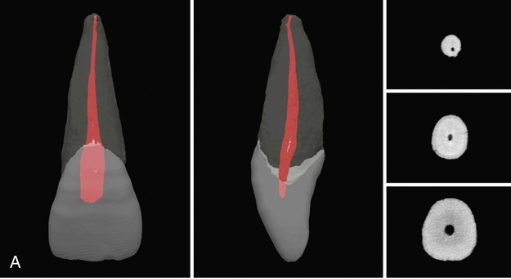
The dental pulp is often referred to as the ___, as opposed to a simple tube or circular space, due to its complexity.
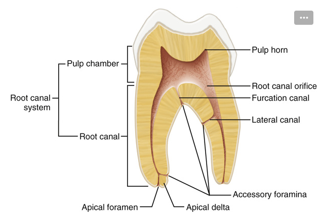
root canal system
Factors such as physiologic aging, pathosis, trauma, and occlusion all can modify the dimensions of the root canal system through the production of ___.
reparative dentin
The root canal system is divided into two portions: the ___, located in the anatomic crown of the tooth, and the ___, found in the anatomic root.
pulp chamber; root canal
The root canal begins as a funnel-shaped canal orifice, generally at or just apical to the cervical line, and ends at the ___, which opens onto the root surface at or within ___ mm of the center of the root apex
apical foramen; 3 mm
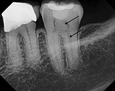
___canals are minute canals that extend in a horizontal, vertical, or lateral direction from the pulp space to the periodontium.
accessory
In ___% of cases, accessory canals are found in the apical third of the root, in ___% in the middle third, and in ___% in the cervical third.
74%, 11%, 15%
Accessory canals that are present in the bifurcation or trifurcation of multirooted teeth are referred to as ___.
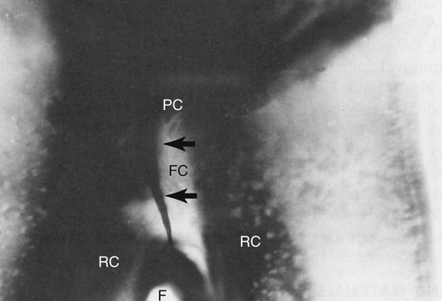
furcation canals
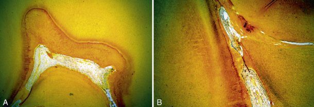
Accessory canals occur in three distinct patterns in the mandibular first molars. A, In 13% a single furcation canal extends from the ___ to the ___. B, In 23% a lateral canal extends from the ___ of a major root canal to the ___. C, About 10% have both ___ and ___canals.
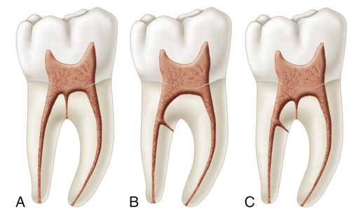
- pulp chamber; intraradicular region
- coronal third; furcation region
- lateral; furcation
Foramina on both the pulp chamber floor and the furcation surface were found in ___% of maxillary first molars, ___% of maxillary second molars, ___% of mandibular first molars, and % of mandibular second molars.
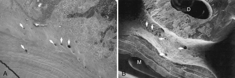
36%; 12%; 32%; 24%
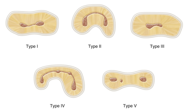
Type I canal: ___
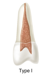
A single canal extends from the pulp chamber to the apex (1).
Type II canal: ___
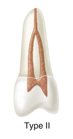
Two separate canals leave the pulp chamber and join short of the apex to form one canal (2-1).
Type III canal: ___
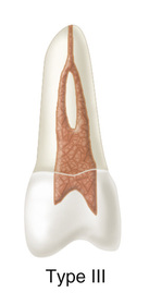
One canal leaves the pulp chamber and divides into two in the root; the two then merge to exit as one canal (1-2-1)
Type IV canal: ___
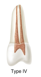
Two separate, distinct canals extend from the pulp chamber to the apex (2).
Type V canal: ___
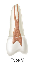
One canal leaves the pulp chamber and divides short of the apex into two separate, distinct canals with separate apical foramina (1-2).
Type VI canal: ___
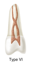
Two separate canals leave the pulp chamber, merge in the body of the root, and separate short of the apex to exit as two distinct canals (2-1-2).
Type VII canal: ___
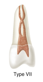
One canal leaves the pulp chamber, divides and then rejoins in the body of the root, and finally separates into two distinct canals short of the apex (1-2-1-2).
Type VIII canal: ___
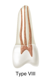
Three separate, distinct canals extend from the pulp chamber to the apex (3).
The only tooth that showed all eight possible canal configurations was the ___.
maxillary second premolar
One well-recognized ethnic variant of root canal anatomy is the higher incidence of ___ in Native American and Asians populations
single-rooted, C-shaped mandibular second molars
It is important to note that if only one canal is present, it usually is located in the ___.
center of the access preparation
If only one orifice is found and it is not in the center of the root, another orifice probably exists, and the clinician should search for it on the ___.
opposite side
In a mandibular second molar, if two orifices are not directly in the mesiodistal midline, a search should be made for another canal on the ___.
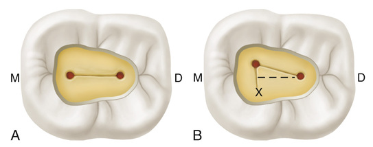
opposite side
Isthmuses are found in 15% of anterior teeth; in maxillary premolar teeth, they are found 52% at the ___ mm level, which puts them primarily in the middle third of the canal
6 mm
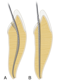
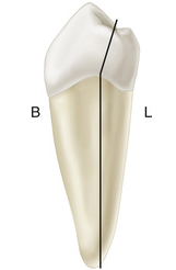
Whenever a root contains two canals that join to form one, the ___ canal generally is the one with direct access to the apex.
lingual/palatal
The classic concept of apical root anatomy is based on three anatomic and histologic landmarks in the apical region of a root:
- ___
- ___
- ___
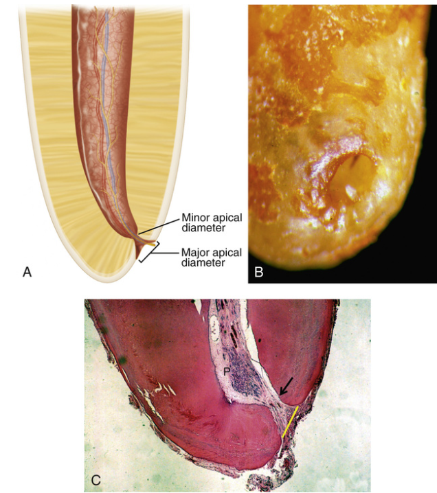
- apical constriction (AC)
- cementodentinal junction (CDJ)
- apical foramen (AF)
Kuttler's description of the anatomy of the root apex has the root canal tapering from the canal orifice to the AC, which generally is ___ mm coronal to the AF
0.5 to 1.5 mm
The ___ generally is considered the part of the root canal with the smallest diameter; it also is the reference point clinicians use most often as the apical termination for enlarging, shaping, cleaning, disinfecting, and filling.
apical constriction
The ___ is the point in the canal where cementum meets dentin; it is also the point where pulp tissue ends and periodontal tissues begin.
cementodentinal junction (CDJ)
Locating the AC and AF is difficult clinically; for this reason, some researchers contend that the ___ is a more reliable reference point.
radiographic apex
Studies have shown that in these cases, a better success rate is achieved when therapy ends at or within ___ mm of the radiographic apex.
2 mm
The objectives of access cavity preparation are to:
- ___
- ___
- ___
- ___
- ___
- ___
- remove all caries when present
- conserve sound tooth structure
- unroof the pulp chamber completely
- remove all coronal pulp tissue
- locate all root canal orifices
- achieve straight- or direct-line access
Krasner and Rankow found that the ___ was the most important anatomic landmark for determining the location of pulp chambers and root canal orifices.
cementoenamel junction (CEJ)
The floor of the pulp chamber is always located in the ___ of the tooth at the level of the CEJ.
center (centrality)
The walls of the pulp chamber are always ___ to the external surface of the tooth at the level of the CEJ; that is, the external root surface anatomy reflects the internal pulp chamber anatomy.
concentric (concentricity)
The distance from the external surface of the clinical crown to the wall of the pulp chamber is the same throughout the circumference of the tooth at the level of the ___.
cementoenamel junction (CEJ)
Except for the maxillary molars, canal orifices are equidistant from a line drawn in a ___ direction through the center of the pulp chamber floor.
mesiodistal (symmetry)
Except for the maxillary molars, canal orifices lie on a line ___ to a line drawn in a mesiodistal direction across the center of the pulp chamber floor.
perpendicular (symmetry)
The pulp chamber floor is always ___ in color than the walls.
darker (color change)
The orifices of the root canals are always located at the junction of the ___ and the ___.
walls; floor (orifice location)
Access cavities on anterior teeth usually are prepared through the ___ tooth surface, and those on posterior teeth are prepared through the ___ surface.
lingual; occlusal
Some authors have recommended that the traditional anterior access for ___ be moved from the lingual surface to the incisal surface in selected cases; this allows better access to the lingual canal and improves canal débridement
mandibular incisors
Removal of all ___ before entering the root canal system is essential to determine restorability.
defective restorations and caries.
A minimum ___ mm of temporary filling material (e.g., Cavit) is needed to provide an adequate coronal seal for a short time.
3.5 mm
___ burs are used extensively in the preparation of access cavities. They are used to excavate caries and to create the initial external outline shape.
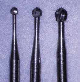
round carbide
Fissure carbide and diamond burs with ___ are safer choices for axial wall extensions.
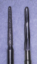
safety tips (non-cutting ends)
___ burs are needed when the access must be made through porcelain or metalloceramic restorations.
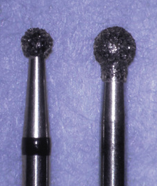
round diamond
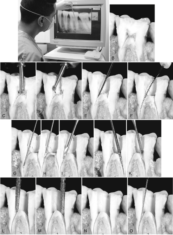
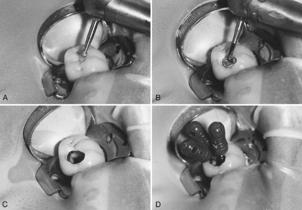
___ burs, such as the Mueller bur or Extendo Bur, can be used to locate a receded pulp chamber or calcified orifice.
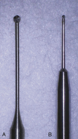
xtended-shank round
The ___ or ___ endodontic explorers are used to identify canal orifices and to determine canal angulation.
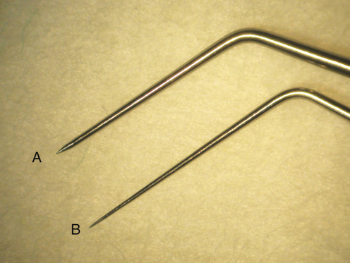
DG-16; JW-17
A sharp ___, which comes in different sizes, can be used to remove coronal pulp and carious dentin.
endodontic spoon
A ___ operative explorer is useful for detecting any remaining overhang from the pulp chamber roof.
#17
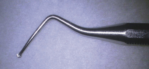
___ can be used to trough and deepen developmental grooves, remove tissue, and explore for canals and provide outstanding visibility compared with traditional handpiece heads.
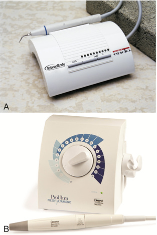
ultrasonic tips
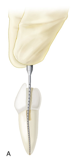
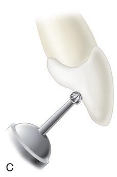
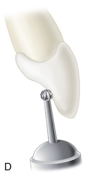
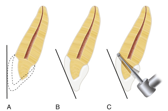
During access preparation for an anterior tooth, cutting commences at the ___ of the anatomic crown.
center of the lingual or palatal surface
During access preparation for an anterior tooth, an outline form is developed that is similar in geometry to an ideal access shape for the particular anterior tooth; it is ___ the projected final size of the access cavity.
half to three quarters
During access preparation for an anterior tooth, the bur is directed perpendicular to the ___ as the external outline opening is created.
lingual surface
During access preparation for an anterior tooth, the angle of the bur is rotated from perpendicular to the lingual/palatal surface to parallel to the ___.
long axis of the root
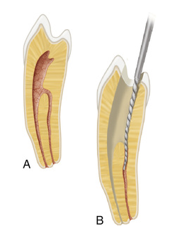
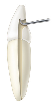
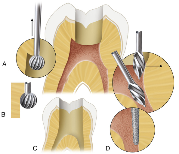
During access preparation for an anterior tooth, once the pulp chamber has been penetrated, the remaining roof is removed by ___.
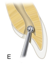
catching the end of a round bur under the lip of the dentin roof and cutting on the bur's withdrawal stroke
During access preparation for an anterior tooth, once the orifice or orifices have been identified and confirmed, the ___ is removed.
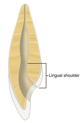
lingual shoulder
During access preparation for an anterior tooth, when a fine, safety-tip diamond bur is used, it should be placed so as to avoid ___.
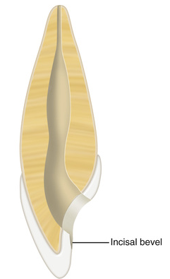
putting a bevel on the incisal edge of the access preparation
Deflected instruments function under more stress than those with minimal or no deflection pressure and are more susceptible to ___.
separation during enlargement and shaping
Without straight-line access, ___ may occur.
procedural errors (e.g., ledging, transportation, and zipping)
During access preparation for an anterior tooth, if the lingual shoulder has been removed properly and a file still binds on the incisal edge, the access cavity should be ___ until the file is not deflected.
extended further incisally
In maxillary premolars, the point of entry that determines the external outline form is on the ___ between the ___.
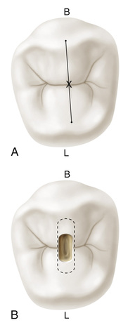
central groove; cusp tips
Crowns of mandibular premolars are tilted ___ relative to their roots; therefore, the starting location must be adjusted to compensate for this tilt.
lingually
In mandibular first premolars, the starting location is halfway up the ___ incline of the ___ cusp on a line connecting the cusp tips.
lingual; buccal
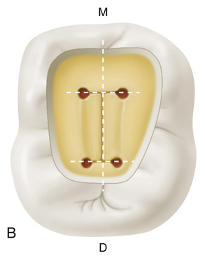
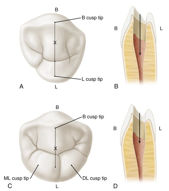
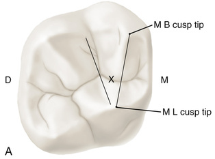
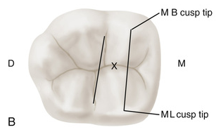
The mesial boundary for molar access cavity preparations is a line connecting the ___. Pulp chambers are rarely found mesial to this imaginary line
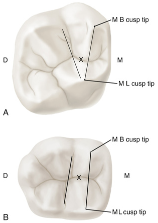
mesial cusp tips
Evaluation of ___ radiographs is an accurate method of assessing the mesiodistal extensions of the pulp chamber.
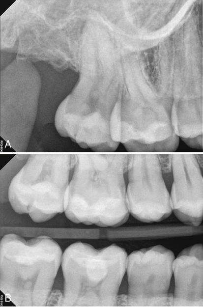
bite-wing
For molar access cavity preparations, a good initial distal boundary for maxillary molars is the ___.
oblique ridge
For molar access cavity preparations, the initial distal boundary for mandibular molars, is ___.
a line connecting the buccal and lingual grooves
For molar access cavity preparations, the correct starting location is on the ___ halfway between the ___.
central groove; mesial and distal boundaries
For molar access cavity preparations, the penetration angle should be toward the ___. Therefore, in maxillary molars, the penetration angle is toward the ___, and in mandibular molars, it is toward the ___.
largest canal; palatal orifice; distal orifice
For molar access cavity preparations, the goal is to funnel the ___ directly into the___.
corners of the access cavity; orifices
For molar access cavity preparations, a ___ bur can be set on the pulp floor and the entire axial wall is shaped at one time, with little or no apical pressure.
safety-tip
For molar access cavity preparations, ideally, the orifices are located at the ___ to facilitate all of the root canal procedures.
corners of the final preparation
For molar access cavity preparations, extension of an orifice into the axial wall creates a ___ effect, which impedes straight-line access.
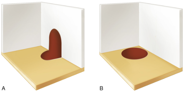
“mouse hole”
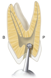
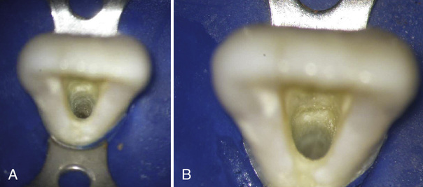
The ___ are shelves of dentin that frequently overhang orifices in posterior teeth, restricting access into root canals and accentuating existing canal curvatures. They can be removed safely with burs or ultrasonic instruments.
cervical bulges
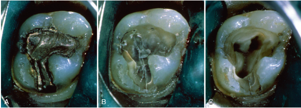
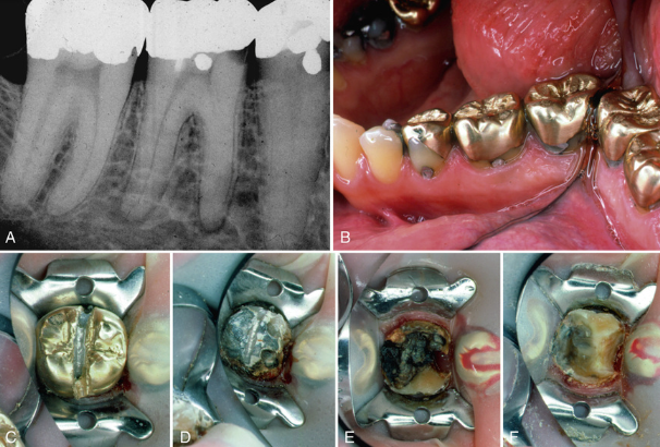
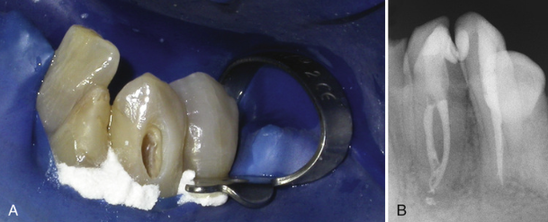
In older teeth with previous caries or large restorations, the pulp chambers typically have ___.
receded or calcified
When treating teeth with loss of significant coronal anatomy, access often is started without a dental dam in place so that ___.
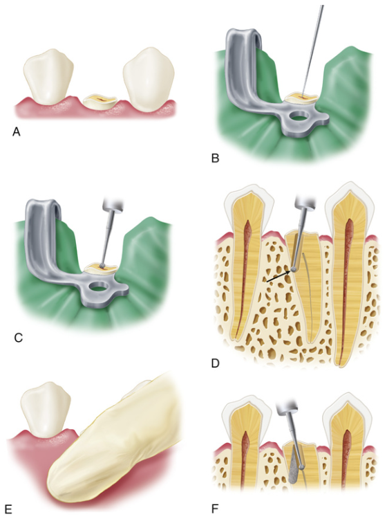
root eminences can be visualized and palpated
When treating previously restored teeth, access may be less predictable because ___.
crown-to-root angulation often is altered when large restorations or crowns correct occlusal discrepancies
Clinicians are ___% more likely to miss these anomalies such as recurrent caries and fracture lines when restorations are not removed completely.
40%
Creating an access through porcelain or metalloceramic restorations must be handled delicately to minimize the potential for ___.
fracture
In patients with crowded teeth, a ___ preparation may be the treatment of choice based on straight-line access principles and conservation of tooth structure.
buccal access
The most common apical canal configuration for the maxillary central incisor is ___.
one canal (100%)
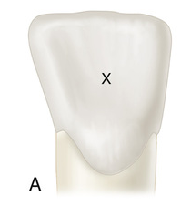
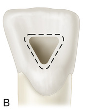
The external access outline form for the maxillary central incisor is a ___.
rounded triangle
The most common apical canal configuration for the maxillary lateral incisor is ___.
one canal (100%)
The external access outline form for the maxillary lateral incisor may be a ___ or an ___.
rounded triangle; oval
The maxillary lateral incisor often has anomalies. One such variation in form is the presence of a ___.
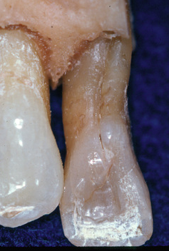
palatal radicular or developmental groove
The most common apical canal configuration for the maxillary canine is ___.
one canal (100%)
The external access outline form for the maxillary canine is ___ or ___ because no mesial or distal pulp horns are present
oval; slot-shaped
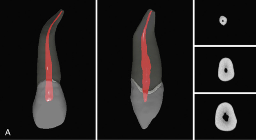
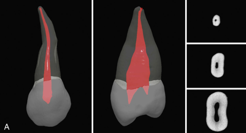
The most common apical canal configuration for the maxillary first premolar is ___.
two canals (69%)
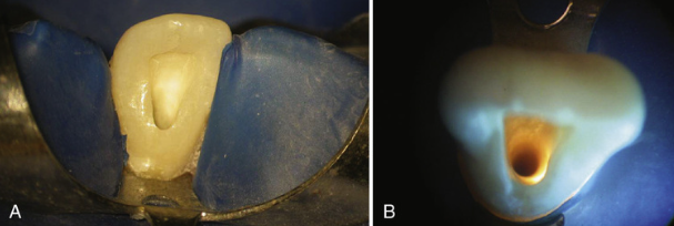
The external access outline form for the maxillary first premolar is ___ or ___.
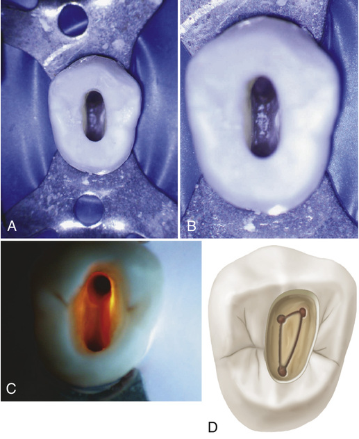
oval; slot-shaped
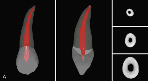
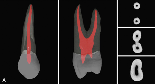
The most common apical canal configuration for the maxillary second premolar is ___.
one canal (75%)
The external access outline form for the maxillary second premolar is ___ or ___.
oval; slot-shaped
The ___ is the largest tooth in volume and one of the most complex in root and canal anatomy.
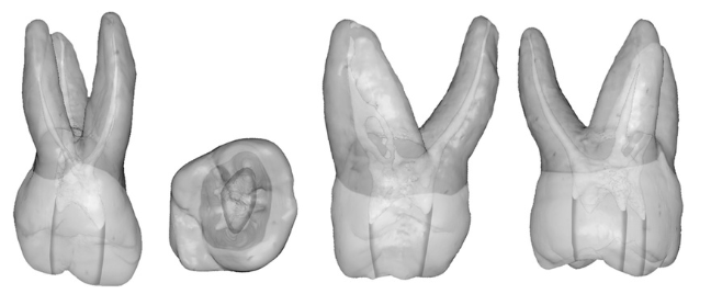
maxillary first molar
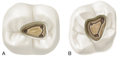
A line drawn to connect the three main canal orifices of a maxillary molar—the mesiobuccal orifice, distobuccal orifice, and palatal orifice—forms a triangle, known as the ___.
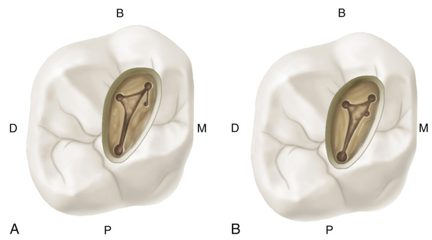
molar triangle
The ___ of the maxillary first molar is the longest, has the largest diameter, and generally offers the easiest access.
palatal root
The most common apical canal configuration for the palatal root of the maxillary first molar is ___.
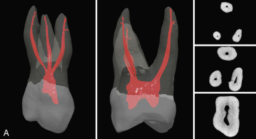
one canal (100%)
The most common apical canal configuration for the distobuccal root of the maxillary first molar is ___.
one canal (100%)
The most common apical canal configuration for the mesiobuccal root of the maxillary first molar is ___.
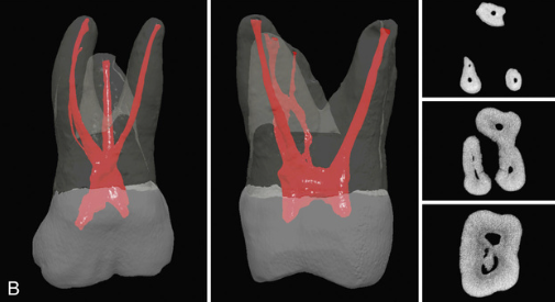
one canal (82%)
Because the maxillary first molar almost always has four canals, the access cavity has a ___ shape.
rhomboid
The most common apical canal configuration for the palatal root of the maxillary second molar is ___.
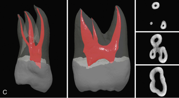
one canal (100%)
The most common apical canal configuration for the distobuccal root of the maxillary second molar is ___.
one canal (100%)
The most common apical canal configuration for the mesiobuccal root of the maxillary second molar is ___.
one canal (88%)
The external access outline form for the maxillary second molar is ___ if there are four canals, ___ if there are three canals, and ___ if there are two canals.
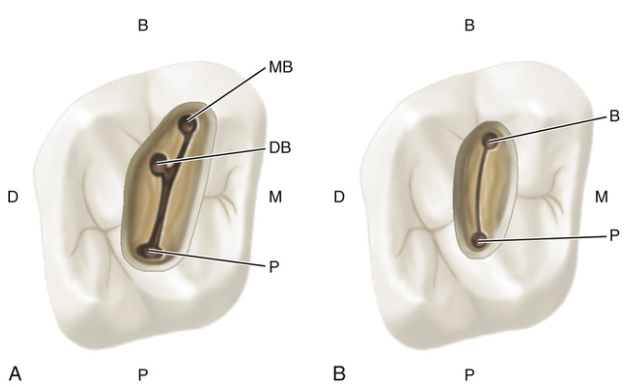
rhomboidal; rounded triangle; oval
The most common apical canal configuration for mandibular incisors is ___.
one canal (97%)
The external outline form for mandibular incisors may be ___ or____, depending on the prominence of the mesial and distal pulp horns
triangular; oval
The most common apical canal configuration for the mandibular canine is ___.
one canal (94%)
The external access outline form for the mandibular canine is ___ or ___ because no mesial or distal pulp horns are present
oval; slot-shaped
As a group, the ___ teeth present anatomic challenges because of the extreme variations in their root canal morphology.
mandibular premolar
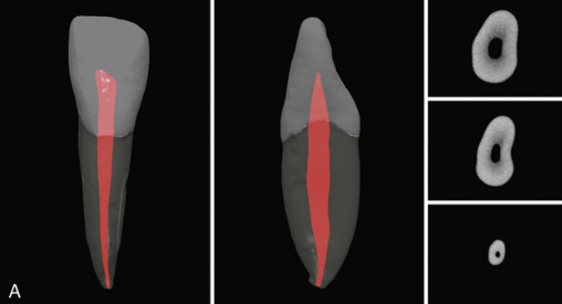
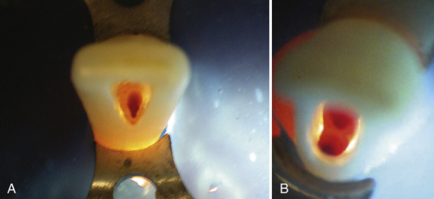
The most common apical canal configuration for the mandibular first premolar is ___.
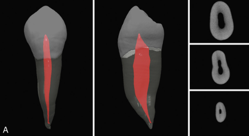
one canal (74%)
The external outline form of the mandibular first premolar is ___ shaped.
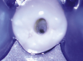
oval
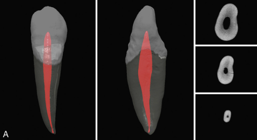
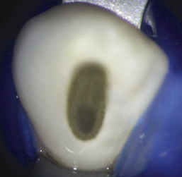
The most common apical canal configuration for the mandibular second premolar is ___.
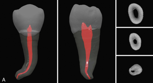
one canal (92.5%)
The external outline form of the mandibular first premolar is ___ shaped.
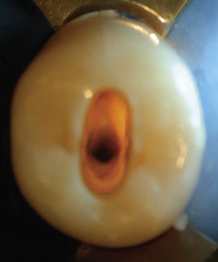
oval
Being the earliest permanent posterior tooth to erupt, the ___ seems to be the tooth that most often requires an endodontic procedure.
mandibular first molar
The most common apical canal configuration for the mesial root of the mandibular first molar is ___.
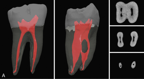
two canals (59%)
The most common apical canal configuration for the distal root of the mandibular first molar is ___.
one canal (85%)
A middle mesial (MM) canal is sometimes present in the developmental groove between the mesiobuccal and mesiolingual canals of the mandibular first molar. The incidence of an MM canal ranges from ___ to ___ %.
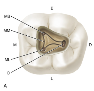
1% to 15%
The access cavity for the mandibular first molar typically is ___ or___, regardless of the number of canals present.
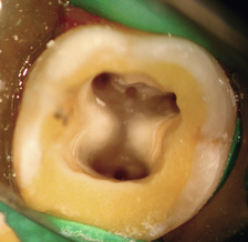
trapezoid; rhomboid
The most common apical canal configuration for the mesial root of the mandibular second molar is ___.
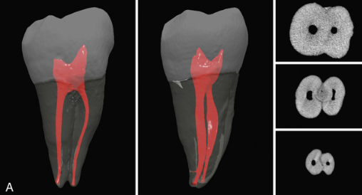
one canal (65%)
The most common apical canal configuration for the distal root of the mandibular second molar is ___.
one canal (95%)
The external access outline form for the mandibular second molar is ___ if there are three canals, ___ if there are two canals, and ___ if there is one canal.
triangular; rectangular; oval
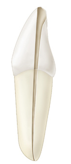
The main cause for C-shaped roots and canals is the ___ on either the buccal or lingual root surface.
failure of Hertwig's epithelial root sheath to fuse
Category I (C1) C-shaped canal: ___.
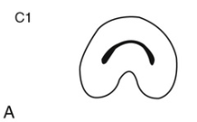
The shape is an uninterrupted “C” with no separation or division.
Category II (C2) C-shaped canal: ___
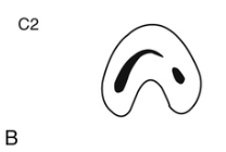
The canal shape resembles a semicolon resulting from a discontinuation of the “C” outline, but either angle α or β should be no less than 60 degrees
Category III (C3) C-shaped canal: ___
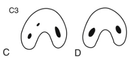
Two or three separate canals and both angles, α and β, are less than 60 degrees.
Category IV (C4) C-shaped canal: ___
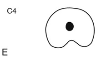
Only one round or oval canal is in the cross-section.
Category V (C5) C-shaped canal: ___
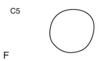
No canal lumen can be observed (is usually seen near the apex only).